It was about five months ago that I started my PhD in the BIOS Lab on a Chip group at the University of Twente. Our group works on a wide variety of topics in the realm of micro- and nanofluidics. Within this interdisciplinary group, I focus on adapting electrode designs in 3D vessels-on-chip to allow for TEER determination in these vessels.
It was during my Bachelor’s in Health Science and Technology at ETH Zurich in Switzerland, that I got introduced to organs-on-chip. The topic immediately peaked my interest. Fast forward a few years, I was doing my Master thesis on measuring the transendothelial electrical resistance (TEER) in a blood-brain barrier-on-chip here at BIOS, where I first heard of NOCI. And of course, I was more than happy when I was offered a PhD position to continue working on a similar topic.
Organs-on-chip have gained a lot of popularity in the last decade. They allow us to better mimic the physiological environment by applying controlled stimuli to the cells. Additionally, human cells can be incorporated which presents an advantage over animal studies. However, with the advancement of organs-on chip arises a need for new adapted monitoring tools. Ideally, such a monitoring tool allows for continuous measurements while being label-free. One tool that can fulfill these requirements is electrical impedance spectroscopy, which is used to measure the electrical resistance over cell barriers such as the wall of a blood vessel. To do this, an electric current is applied between electrodes on the inside and the outside of the vessel, and the resulting voltage drop is measured. From this, the resistance of the vessel wall, the so-called TEER can be calculated using Ohm’s Law. The tighter the cells are connected to each other through tight junctions, the more they restrict the flow of ions across the vessel wall, which increases the TEER.
The expected TEER depends on the type of vessel that is being investigated. For example, the blood vessels in the brain exhibit a relatively high TEER due to the very tight blood-brain barrier which needs to fine-tune which substances are able to enter the brain. This also restricts the passage of ions that carry the current. However, if there are holes in the cell layer, the TEER will drop. So by measuring the TEER, we can obtain information about the integrity and the tightness and of the barrier. This is a useful tool to monitor vessels during their formation, as well as during drug testing.
TEER measurements are an established technique that has been used for many years and has also been incorporated into some organs-on-chip. However, currently this resistance is mainly measured for 2D cell cultures, either in transwells or in sandwich-design chips. In both cases, important physiological aspects are missing, such as the correct vessel size and curvature, or exposure to shear stress through flow. In my project, I attempt to create a 3D vessel-on-chip, and adapt the electrode design in a way that allows for the determination of the TEER of this vessel.
I’m excited to work on bringing this technique that is currently used for 2D cultures into the third dimension by combining my knowledge of microfabrication, cell cultivation, impedance measurements and computational simulations. I am also really happy to be part of NOCI, which has already given me great opportunities to network and exchange ideas with other researchers working on different aspects of organs-on-chip , and I look forward to all the future discussions and collaborations within this amazing network.
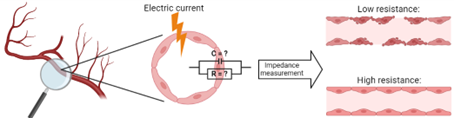
Simplified schematic of the principle of transendothelial electrical resistance (TEER) measurements. By measuring the resistance to electrical current over the cell barrier, insight into the barrier integrity and tightness can be gained. Figure created with BioRender.com.

Muriel A. Holzreuter
NOCI PhD student University of Twente